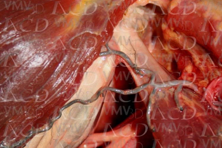
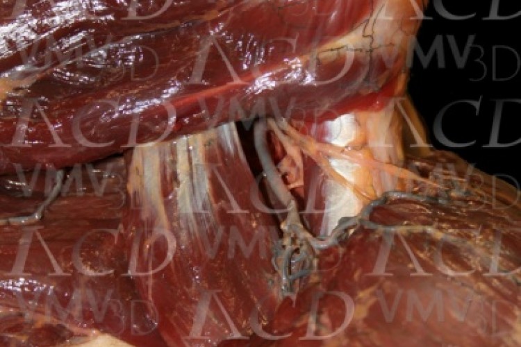
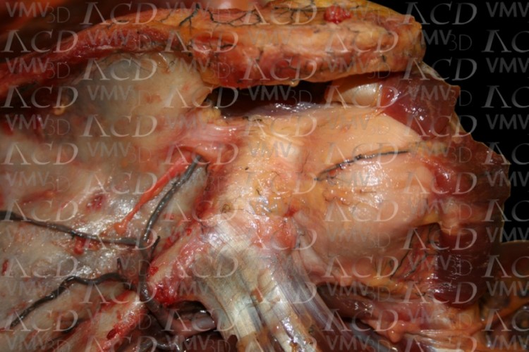
Anterior view of shoulder showing toracoacromial artery and its branches (deltoid and acromial).
Pectoral Girdle vascular-nerves
Anatomical images are displayed on vascular and nerve structures with their relations with the muscles of the shoulder girdle.
Toracoacromial artery.
Anterior view of shoulder showing toracoacromial artery and its branches (deltoid and acromial).
Toracoacromial artery.
Anterior view of shoulder showing toracoacromial artery and its branches (deltoid and acromial).
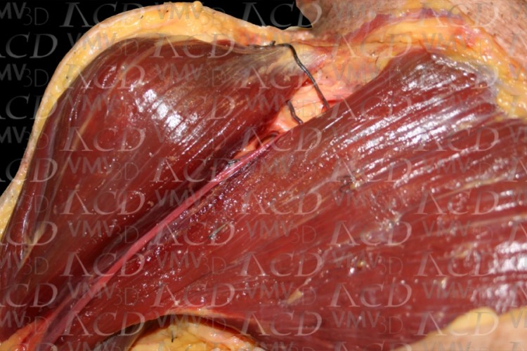
Toracoacromial artery.
Anterior view of shoulder showing toracoacromial artery and its branches (deltoid and acromial).
Axillary nerve and posterior humeral circumflex artery.
Detail of the output of the posterior humeral circumflex artery and axillary nerve through the space of Velpeau.
Notch of the scapula.
Top view of the notch of the scapula showing the passage of the suprascapular artery and nerve.
Notch of the scapula.
Top view of the notch of the scapula showing the passage of the suprascapular artery and nerve.
Notch of the scapula.
Top view of the notch of the scapula showing the passage of the suprascapular artery and nerve.
Axillary nerve and posterior humeral circumflex artery.
Detail of the output of the posterior humeral circumflex artery and axillary nerve through the space of Velpeau.
Suprascapular nerve and artery.
Detail of the passage of the suprascapular artery and nerve in the neck of the scapula, held by the spinoglenoid ligament.
Notch of the scapula.
Top view of the notch of the scapula showing the passage of the suprascapular artery and nerve.
Suprascapular nerve and artery.
Detail of the passage of the suprascapular artery and nerve in the neck of the scapula, held by the spinoglenoid ligament.
Notch of the scapula.
Top view of the notch of the scapula showing the passage of the suprascapular artery and nerve.
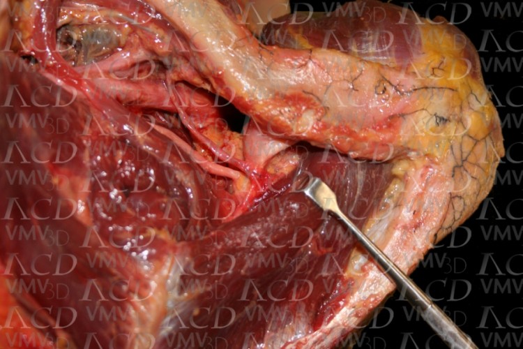
Toracoacromial artery.
Anterior view of shoulder showing toracoacromial artery and its branches (deltoid and acromial).

