Radiology
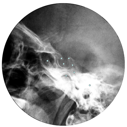
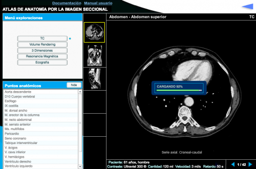
Atlas of Anatomy by Radiological Sectional Image, includes more than 14,000 images taken by the different current techniques of radiology (RX, RNM, TC, Ultrasound, etc.) grouped and sequenced to be able study human anatomy by using the more modern techniques.
| Precio | 50,00 € |

Atlas of Anatomy by the Radiological Sectional Image
Atlas of Anatomy by the Radiological Sectional Image, includes more than 14,000 images taken by different radiology techniques (Rx, RNM, CT, ultrasound, PET, etc.) grouped and sequenced in order to study the human anatomy using techniques most modern radiological. All the images are labeled indicating the name of the different relevant anatomical elements (muscles, bones, arteries, veins, organs, etc.).
Systems and Devices menu:
Thorax: mediastinum-thoracic wall, lung-pleura, tracheobronchial pathway, heart, coronary circulation, thoracic operculum, mammary gland.
Abdomen: upper abdomen, stomach-duodenum, small and large intestine, liver, bile duct, pancreas, peritoneum-peritoneal cavity, retroperitoneum, kidney, urinary tract, male pelvis, female pelvis.
Head and neck: neck, larynx, cavum-pharynx, facial-facial shape, temporomandibular joint, upper jaw and maxilla, paranasal orbit-sinuses, boulder, base of skull, cranial cavity.
Locomotor: cervical spine, dorsal column, lumbosacral spine, sacrum, shoulder, elbow, wrist, hand, hip, knee, ankle, foot.
Vascular: encephalic vessels, neck vessels, supra-aortic trunks, azygos-cava system, pulmonary vessels, thoracic aorta, abdominal aorta, iliac artery, celiac trunk, renal arteries, arteries, mesenteric, upper extremity, femoropopliteal, lower extremity, leg, foot, ilio cava axis, suprahepatic veins, portal system.
Nervous System: brain, cranial nerves, sella turcica, brachial plexus

